Optic neuritis is inflammation or injury affecting the optic nerve, which carries visual information from the eye to the brain. It can cause blurred vision, changes in color perception, or pain when moving the eye. Symptoms deserve prompt attention, especially when vision changes suddenly. This guide explains possible signs and causes, what an evaluation may involve, and how treatment decisions are made.
Contact Abita Eye about an eye evaluation
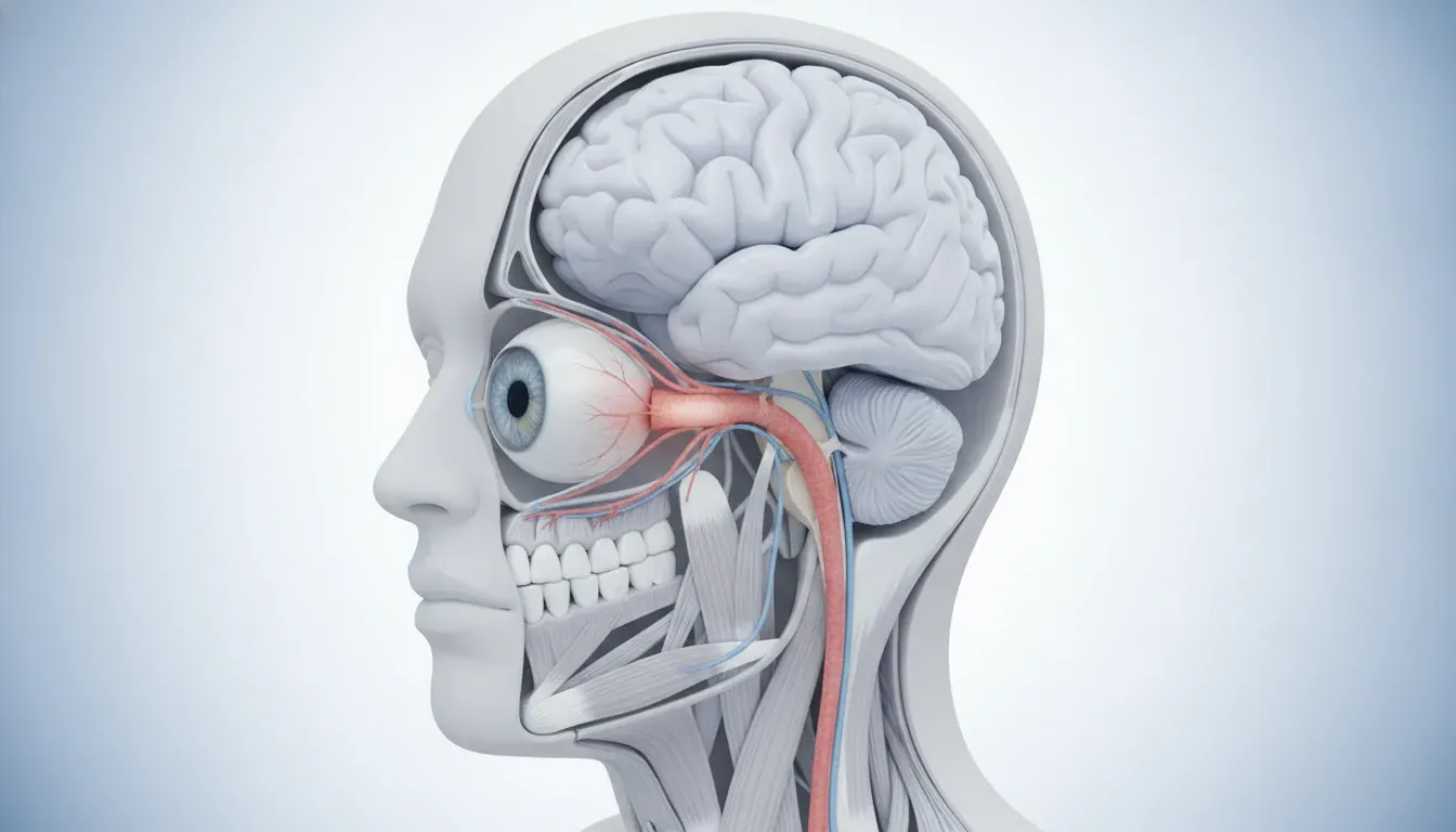
The optic nerve is made of nerve fibers that transmit signals from the retina to the brain. When those fibers become inflamed or otherwise injured, the affected eye may not send visual information normally. Clinicians use the term optic neuritis for inflammation of the optic nerve. The condition can occur on its own or in association with a broader illness, including some immune-system or infectious conditions.
Optic neuritis is one possible explanation for vision symptoms, not a conclusion that can be reached from symptoms alone. Several eye and nervous-system problems can cause blurred or reduced vision, pain, or changes in color. A clinician needs to assess the full pattern and decide whether testing or referral is appropriate. A person should not assume that eye pain or blurred vision is optic neuritis, nor dismiss an abrupt vision change because it does not match a familiar description.
The phrase “optic neuritis” may describe different patterns. Some cases are considered typical based on the person's age, symptoms, examination, and clinical course. Other cases are atypical because they are unusually severe, affect both eyes, recur, or have features that suggest a different underlying cause. These labels are clinical descriptions; they do not establish the cause by themselves.
It helps to separate three questions that are sometimes blended together: Is the optic nerve affected? What caused the problem? What should happen next? An eye examination and selected tests help address the first question. A review of symptoms, medical history, imaging, and sometimes laboratory findings may help answer the second. The third depends on the findings and the person's health, so treatment and follow-up are individualized. Even if a clinician suspects optic neuritis early, the cause may remain uncertain until the evaluation is complete.
Symptoms can develop over hours or days, and their severity varies. Many people notice changes in one eye, but symptoms are not identical for everyone. Possible symptoms include:
Not everyone has eye pain, and pain alone does not confirm optic neuritis. Some people notice the problem only when they cover one eye at a time, because the unaffected eye can compensate during everyday activities. If a new change is suspected, comparing each eye separately may help describe what is happening, but it is not a substitute for an examination.
Symptoms can be subtle at first. A person may describe a foggy patch, a washed-out image, or a difference in brightness rather than “vision loss.” Children may have difficulty explaining these changes. Parents can note observable behaviors, such as closing one eye, turning the head to look at an object, or avoiding visually demanding tasks, and share those observations with a clinician. These behaviors have many possible explanations and do not diagnose optic neuritis.
When describing symptoms, use concrete examples instead of trying to name the condition. For instance, say whether a face looks less clear, whether a red object appears less bright through one eye, or whether a patch of the view seems missing. Mention the time the change was first noticed and whether it has stayed the same, improved, or worsened. If symptoms fluctuate, describe when that happens. These details help a clinician understand the experience without requiring the patient to interpret its cause.
A simple comparison can help organize observations while waiting for professional advice: in a safe, seated setting, look at the same object with one eye covered and then the other. Do not press on the eyes, use bright lights, or delay care to repeat a home check. Write down any obvious difference, but remember that this comparison is not a vision test and cannot rule out a serious problem. If the change is sudden or substantial, seek appropriate care rather than spending time trying to measure it.
New or sudden vision loss should be taken seriously. Contact an eye-care professional or seek urgent medical evaluation promptly for a sudden change in vision, especially if it is worsening, affects one eye substantially, or comes with pain on eye movement. If vision is severely reduced, symptoms are rapidly progressing, or an urgent eye evaluation is not available, seek emergency care. Do not wait for an online article to identify the cause.
Seek emergency medical help for vision changes that occur with other serious symptoms, such as weakness, trouble speaking, confusion, a severe sudden headache, loss of coordination, or facial drooping. Those symptoms can signal a time-sensitive problem that requires immediate assessment. If vision is impaired, avoid driving yourself; ask someone to take you or contact emergency services when appropriate.
These recommendations are about the urgency of assessment, not a diagnosis. Sudden vision changes may have causes other than optic neuritis, some of which also need urgent treatment. When calling a practice, describe when the change began, which eye is affected, whether it is getting worse, and any additional symptoms. Staff can advise on the appropriate next step, but a clinician must determine the cause.
For a call or urgent visit, prepare a short timeline if doing so will not delay care. Note the last time vision seemed normal, when the change was first noticed, whether one or both eyes seem affected, and whether pain or other symptoms appeared before or after it. Mention recent injuries, new medicines, or a significant illness if relevant. A person may not know the exact onset, especially if symptoms started gradually or were first noticed on waking; an approximate time is still useful.
Do not attempt to drive if vision, depth perception, or comfort makes driving unsafe. Arrange a ride, and bring someone who can help explain the timeline if the patient is a child or feels unwell. If emergency symptoms appear while waiting for an appointment, change the plan and seek emergency assessment. Routine scheduling and an online contact form are not substitutes for emergency services when the symptoms are severe.
Optic neuritis can be associated with the body's immune response. In some people, inflammation of the optic nerve occurs in connection with a demyelinating condition. Demyelination means damage to the protective covering around nerve fibers. Multiple sclerosis (MS) is one condition associated with optic neuritis, and an episode may be an early sign that prompts further neurologic evaluation. An episode does not, by itself, prove that a person has MS.
Other immune-mediated conditions can affect the optic nerve. Neuromyelitis optica spectrum disorder (NMOSD) and myelin oligodendrocyte glycoprotein antibody-associated disease (MOGAD) are examples clinicians may consider in particular patterns of illness. These conditions are not interchangeable, and their symptoms and evaluation differ. A clinician decides whether testing for them is relevant based on the person's history, examination, imaging, and course.
Infections and other inflammatory illnesses can also be considered, depending on the person's circumstances. Sometimes no specific cause is identified after evaluation. The range of possibilities is one reason not to start treatment based on a suspected diagnosis or someone else's experience. The right workup depends on what the examination shows and whether there are clues to a broader health condition.
Risk factors and context matter. A clinician may ask about previous episodes, other vision or neurologic symptoms, recent illness, medications, and general health. These questions help shape the evaluation; they do not mean that a particular answer caused the problem. Avoid stopping prescribed medicine or starting supplements or leftover medication without medical advice.
The cause is not always apparent at the first visit. A clinician may initially describe the finding as suspected optic neuritis or an optic nerve problem while arranging additional assessment. That wording reflects what is known at that point, not necessarily an incomplete or mistaken evaluation. Some causes become more or less likely as test results and the clinical course are reviewed. Patients can ask which possibilities are being considered, which findings support them, and what result would change the plan.
It is also useful to distinguish an association from a certainty. A condition may be associated with optic neuritis without being present in every person who develops it. Likewise, having a relevant health condition does not mean that every episode of blurry vision is caused by optic nerve inflammation. A clinician considers the complete picture rather than relying on a single risk factor or symptom. This is why broad internet lists of causes cannot determine an individual's diagnosis.
Many different conditions can affect vision, and some may feel similar at first. Refractive changes can make vision blurry, while problems affecting the cornea, lens, retina, or other parts of the visual pathway can cause dimness, distortion, or missing areas. Headache and eye discomfort can occur for many reasons. The location, timing, and pattern of symptoms help a clinician decide what to examine; no single symptom reliably separates these conditions at home.
Sudden painless loss of vision, flashes or a curtain-like shadow, marked redness, halos, or a severe headache may point toward other urgent eye or health problems. These descriptions are not a self-diagnosis checklist. People may describe symptoms differently, and more than one problem can be present. The safest response to a sudden or significant change is prompt professional assessment rather than trying to match symptoms to a condition online.
Some optic nerve disorders are not inflammatory. A clinician may use broader terms such as optic neuropathy while investigating damage or dysfunction of the optic nerve. The cause may relate to blood flow, pressure, infection, inflammation, medication effects, or another factor. This distinction matters because treatment choices depend on the cause, not simply on the fact that vision is affected.
Tell the clinician about the exact sequence of symptoms. For example, note whether pain began before the vision change, whether colors appear different, whether symptoms involve one or both eyes, and whether there were flashes, floaters, or a field defect. A description is useful even when it is imperfect. Avoid assuming that a symptom rules a condition in or out.
Some patterns may help guide the examination but should not be used to decide at home that a problem is harmless. For example, blur that clears with blinking can have a different set of possible explanations from a persistent dark patch, but the patient cannot reliably locate the cause based on that difference alone. A curtain-like shadow or new flashes and floaters can raise concern for problems elsewhere in the eye, while pain with movement may lead a clinician to assess the optic nerve. These are clues to report, not diagnostic rules.
When searching for information, be cautious about comparing one person's recovery or test results with another's. Similar words such as “blurred,” “dim,” or “painful” can describe very different findings. Even people with the same diagnosis can have different causes, severity, and care plans. The most useful next step is to share specific observations with a clinician and ask what alternatives have been considered.
An evaluation often begins with a careful history and an eye examination. The clinician may ask when the symptoms began, how they have changed, whether one or both eyes are affected, whether there is pain, and whether there have been other symptoms. Bring a current medication list and relevant medical history if possible. If the patient is a child, a parent or caregiver can describe what they have noticed and when.
Vision testing may include measuring how clearly each eye sees, checking pupils, examining color perception, and assessing the visual field. A clinician may look at the optic nerve and retina using instruments that examine the back of the eye. The appearance of the optic nerve can vary; a normal-looking nerve at the front of the eye does not necessarily rule out inflammation farther along the nerve.
Depending on the findings, clinicians may recommend imaging, such as magnetic resonance imaging (MRI) of the brain and/or orbits. Imaging can help assess the optic nerve and look for findings that inform the broader evaluation. It is not the only part of diagnosis, and an imaging result must be interpreted alongside symptoms and examination findings.
Other tests may be considered in selected cases. Optical coherence tomography (OCT) creates detailed images of layers at the back of the eye. Visual field testing maps areas a person can see. Visual evoked potential testing measures how visual signals travel toward the brain. Blood tests, and sometimes other neurologic investigations, may be used when the history or examination raises questions about a specific underlying condition. Not every patient needs every test.
The evaluation may involve more than one kind of clinician. Depending on the findings, an eye-care clinician may coordinate with a neurologist, neuro-ophthalmologist, or other medical specialist. Which professionals are involved depends on the patient's needs and local availability. Ask who will explain test results, what symptoms should prompt a call, and whether a follow-up visit is needed.
Each part of the assessment contributes a different piece of information. Visual acuity describes how clearly a person can read a chart at the time of testing; it does not capture every aspect of vision. Color comparison, pupil responses, and visual field testing can reveal differences that a standard chart may not show. Examination of the eye can help identify problems in structures such as the cornea, lens, retina, and optic nerve head. Together, these observations guide decisions about further tests.
It can help to ask what each proposed test is meant to clarify. Is it checking the structure of the optic nerve, mapping areas of reduced vision, looking for signs of a broader neurologic condition, or investigating a possible infection or immune process? Understanding the purpose makes it easier to follow the plan and know which clinician will interpret the result. The clinician can also explain whether the test is time-sensitive and what to do while waiting.
Some appointments include several tests and may take longer than a routine vision check. Ask whether drops will be used, whether the visit could temporarily affect near vision or light sensitivity, and whether someone should accompany the patient. Requirements differ by test and clinic, so do not assume that every evaluation includes the same procedures. If a test is uncomfortable or difficult, tell the staff; they can explain the steps and discuss appropriate accommodations.
For a child, a clinician can adapt the examination to the child's age and ability to participate. A caregiver can bring glasses, prior prescription information, and records from earlier evaluations when available. If the child cannot complete a particular test, the care team may consider other ways to assess vision. An incomplete home check should not delay an evaluation when a new vision change is present.
Tests answer different questions. A visual field test records which areas a person can see during the test. OCT can document the structure of retinal layers that include the fibers connected with the optic nerve. MRI can show features that help clinicians assess the optic nerve and, when appropriate, the brain. None of these results should be interpreted in isolation or as a substitute for the patient's symptoms and examination.
A test can be useful even when it does not provide a simple yes-or-no answer. The clinician may compare results over time, look for a pattern, or use testing to assess another possible explanation. Some findings may be nonspecific. If you receive a report before speaking with the clinician, ask what the finding means in your particular case rather than relying on a phrase from the report or an online search.
Ask who will communicate results and when. If several clinicians are involved, clarify which person is coordinating the plan and where questions should go. If a test or referral is recommended but difficult to arrange, tell the care team; they may be able to explain alternatives or the urgency. A test plan is individualized, and a person should not try to order or interpret investigations independently.
When reviewing a result, useful questions include: What did this test show? What did it not show? Does the result change the suspected cause or treatment? Is another test needed, and what is its timing? A normal result in one part of an evaluation does not necessarily explain all symptoms or exclude every possible condition. The clinician can put the result in context and explain whether monitoring is still needed.
Keep copies of reports when several specialists are involved, and confirm that each clinician has access to the relevant information. If a result is pending, ask how it will be delivered and what to do if no update arrives by the expected time. Do not change medication or cancel follow-up based only on a brief report label. Decisions should reflect the whole evaluation and the patient's current symptoms.
Treatment depends on the likely cause, the pattern and severity of symptoms, test results, and the person's overall health. There is no single treatment plan that is right for every case. Clinicians may monitor some situations, recommend treatment for inflammation, investigate an underlying condition, or coordinate care with another specialist.
Corticosteroids may be considered in some cases. They can affect the course of an acute episode, but the decision to use them, and the route and dose, are medical decisions. The potential benefits and risks need to be weighed for the individual. Steroids can have side effects and may be unsuitable in some circumstances. Do not use leftover prescription medicine, borrow medication, or change a prescribed dose without speaking with the treating clinician.
When optic neuritis is linked to a specific underlying disorder, treatment may also address that disorder. The approach can differ among conditions, so the cause matters. A clinician may recommend additional testing or a specialist consultation before deciding on longer-term care. Treatment directed at one type of optic neuritis should not be assumed to work for another.
Some symptoms improve over time, but the timing and degree of recovery vary. People may continue to notice changes in color, contrast, or clarity even as other aspects of vision improve. A clinician can monitor vision and discuss practical adjustments during recovery. Never interpret improvement as proof that the original problem was harmless or that follow-up is unnecessary if it was recommended.
It is reasonable to ask about the goal of any recommended treatment. Is the aim to address an active episode, treat an identified underlying condition, reduce a particular risk, or support function while vision changes? Ask how the clinician will decide whether the plan is working and which side effects or new symptoms should be reported. The answers will depend on the suspected cause and the treatment selected.
Before beginning a medicine, tell the prescriber about other medicines, supplements, allergies, and health conditions. Ask for written instructions if the schedule is complex, and check whom to contact about a missed dose or possible side effect. Do not stop a course early or extend it on your own. If treatment is being considered while more tests are pending, ask how those results affect the decision and when the plan will be reviewed.
Monitoring is also an active plan, not necessarily a sign that symptoms are being ignored. A clinician may recommend repeat vision measurements or another visit to see whether findings are changing. Ask what interval is appropriate, what changes should prompt an earlier call, and where to seek care outside office hours. If symptoms become much worse before the planned check, contact the care team promptly or use urgent care as appropriate.
Recovery is different for each person. Symptoms may change over days or weeks, and the final visual result cannot be predicted from a short description alone. The affected eye may remain less sharp or perceive color and brightness differently. Some people have a single episode; others may have recurring symptoms or an underlying condition that requires continuing care.
Follow the clinician's instructions about reassessment and tests. Keep a brief record of meaningful changes, including which eye is affected and when symptoms change. If vision worsens, a new symptom appears, or the person develops a concerning neurologic problem, contact a clinician promptly or seek urgent care as appropriate. Do not wait for a scheduled follow-up if the situation has changed significantly.
During recovery, everyday tasks may need adjustment. Use good lighting, enlarge text on screens, and take breaks if reading is uncomfortable. If vision is too limited for safe driving or work, ask a clinician about restrictions and support. These practical measures do not treat inflammation, but they can help a person manage visual difficulty while receiving care.
Recovery is not always a straight line. A person may notice improvement in one area, such as overall clarity, while color or contrast still seems different. Fatigue, lighting, and the visual demands of a task can make symptoms more noticeable, but a change should not automatically be attributed to tiredness. Record a meaningful worsening and ask the care team whether it warrants earlier assessment. Sudden or marked deterioration should be treated as urgent.
Consider practical adjustments at home and work while vision is affected. Increase contrast on a screen, enlarge text, reduce glare, and place frequently used items where they are easy to find. For reading, try shorter sessions with breaks rather than pushing through discomfort. If one eye sees differently, give yourself more time with stairs, pouring liquids, or other tasks that depend on depth perception. These are temporary supports, not a replacement for medical care.
Driving decisions should be based on safe vision and clinician guidance, not simply on whether a person can read a familiar sign. If the view is dim, distorted, or difficult to judge, avoid driving until it has been assessed. Ask about work, school, sports, or other activities that may need modification. A written note describing any recommended restrictions can help a patient explain temporary needs to an employer or school.
Follow-up can also help identify whether the underlying cause needs longer-term attention. Ask if the care team expects the episode to be isolated, whether a specialist will review the findings, and what symptoms should lead to a new assessment. Keep scheduled visits even if vision feels better unless the clinician advises otherwise. Improvement is encouraging, but it does not by itself answer what caused the episode.
No. Optic neuritis describes inflammation or injury involving the optic nerve. MS is a disease of the central nervous system. Optic neuritis can occur in people with MS, and an episode may lead clinicians to look for evidence of MS, but many other explanations are possible. A diagnosis requires a complete medical assessment rather than one symptom or one test in isolation.
If MS is a concern, the clinician may discuss imaging, neurologic symptoms, examination findings, and whether additional testing is appropriate. Ask the clinician to explain what is known, what remains uncertain, and what the next step is. It is reasonable to ask whether the evaluation is assessing the optic nerve alone or also looking for a broader neurologic cause.
It can be stressful to hear that a vision problem may lead to a neurologic workup. A workup is a process of gathering information, not a confirmation of a particular diagnosis. Ask what findings prompted the recommendation, when results are expected, and whether a neurologist or neuro-ophthalmologist will be involved. If more than one possibility remains, the clinician can explain what would support or weaken each one and what follow-up is planned.
Children can develop optic nerve problems, but they may not describe symptoms in the same way as adults. A child might say that one eye is blurry, colors look strange, or an image is hard to see. Younger children may not have the words to explain a change, so caregivers may notice that the child covers one eye, sits closer to a screen, turns the head, or has trouble with familiar visual tasks. These signs can have many causes and do not confirm optic neuritis.
Prompt assessment is important when a child has a new vision change, particularly if it is sudden or getting worse. Share when the behavior or symptom began, whether it seems to affect one eye, and whether there is pain or another health concern. The clinician can select age-appropriate tests and determine if specialist input is needed. Do not try to test a child by relying only on an online chart or by comparing their behavior with another child's.
Caregivers can help by offering calm, concrete descriptions and bringing the child's medical and medication history. If the child has difficulty explaining what is wrong, avoid repeatedly asking leading questions; note the child's own words and observed behavior. Follow the clinician's guidance about school, screens, activities, and reassessment. Any sudden or severe loss of vision in a child should be treated as urgent.
Before the visit, caregivers can write down when the change was first noticed and what the child was doing at the time. Note whether the child mentions one eye, avoids certain tasks, bumps into objects, or has difficulty recognizing details at a distance. Ask teachers or other caregivers whether they have noticed a change, but treat those observations as context rather than proof of a diagnosis. If the child uses glasses, bring them and describe whether the problem is different with them on.
Children may feel worried if they hear unfamiliar medical terms or see several tests being prepared. A brief, simple explanation such as “the clinician will check how each eye sees” may be more reassuring than speculating about a cause. Allow the care team to explain the examination in age-appropriate language. If the child cannot complete a test, ask whether another approach is possible and whether the limitation affects the next step.
Clear details can help the care team understand the problem. Before an appointment, write down the approximate time symptoms started and whether they came on suddenly or gradually. Note whether symptoms affect one eye or both, what the vision looks or feels like, whether there is pain with eye movement, and whether anything has changed since onset.
If symptoms are severe or rapidly worsening, do not delay urgent care to gather records. A family member or caregiver may help explain what has changed, particularly for a young child or someone whose vision is substantially affected. The clinician can advise what information is useful after immediate needs are addressed.
It may help to bring a short written summary rather than trying to remember every detail during a stressful visit. Include the main concern in one sentence, the timeline, the eye or eyes involved, and any related symptoms. List current medicines by name and dose when known, but do not postpone evaluation to create a perfect list. Mention any recent change in medication or health that may be relevant.
Consider taking a support person if you expect to have difficulty seeing paperwork, remembering instructions, or traveling afterward. Ask permission before recording instructions; instead, a written summary or patient handout may be available. Before leaving, repeat back the next steps: which test or referral is planned, when to expect results, whom to contact with questions, and what symptoms mean you should seek help sooner.
If a referral is recommended, ask whether the referring clinician will send the examination findings and test records, or whether you should request copies. Confirm how to contact the receiving office and whether you need to arrange the visit yourself. If you cannot obtain the recommended appointment in the expected timeframe, tell the referring care team so they can clarify the urgency or discuss another route. Do not assume that silence means the referral was completed.
Optic neuritis may require a different care pathway from a routine vision concern. An initial eye evaluation can help determine what is happening and whether further assessment is needed. The right clinician and location depend on the symptoms and examination; do not assume that a particular service is offered at every office or that an online description confirms availability for a specific condition.
Abita Eye Group provides general and pediatric ophthalmology care. The practice also describes optometry and full optical services at many locations; Medical Retina and Oculoplastics are specifically offered at the Spring Hill office. These service descriptions do not establish that every location evaluates optic neuritis or that a particular appointment type is suitable. When contacting the practice, explain that there is a new vision change and ask whether the office can guide you to an appropriate evaluation. For sudden or serious symptoms, seek urgent medical care rather than waiting for a routine appointment.
Review the services information and office locations, then contact the practice with questions about the appropriate next step. The pages can help with general practice information, but a clinician must determine which evaluation is appropriate for an individual.
When contacting an office, be concise about the reason for calling: “I have a new change in vision that began [time], and it affects [one/both] eyes.” Mention pain or rapidly worsening symptoms and ask whether the office can advise on an appropriate next step. Do not describe a suspected diagnosis as confirmed. If the office cannot assess the concern promptly, ask where to seek urgent care; use emergency services for severe or rapidly progressing symptoms.
For a non-emergency inquiry, clarify what the appointment will cover and whether any outside records should be sent in advance. Ask who will review the findings and how to follow up about tests. Service pages and office listings provide general practice information, but appointment suitability and clinician availability need confirmation with the practice. Patients should not wait for a routine booking if their symptoms become urgent.
Contact Abita Eye to ask about an evaluation
It can, although one-eye symptoms are common in some patterns of optic neuritis. Symptoms affecting both eyes, particularly when severe or unusual, make a careful evaluation important. Bilateral symptoms do not establish a specific cause, so clinicians consider the whole history and examination.
No. Pain with eye movement can occur with optic neuritis, but it can also have other causes, and some people with optic neuritis do not have pain. New pain with a vision change should be assessed by a clinician rather than diagnosed from that symptom alone.
Yes. Sometimes evaluation does not identify a specific underlying cause. In other cases, testing or follow-up may clarify an association. A clinician can explain what has and has not been established and whether monitoring or more tests are appropriate.
Recovery varies, and no general article can predict an individual's outcome. Some people improve substantially, while others notice lasting differences in sharpness, color, or contrast. Follow-up allows the care team to assess changes and discuss support based on the person's vision.
Do not wait when vision loss is sudden, worsening, or accompanied by significant pain or other concerning symptoms. Seek prompt professional advice; use emergency care for severe or rapidly progressing symptoms or when urgent assessment is otherwise needed. A clinician can determine the cause and the appropriate response.
Optic neuritis is only one possible cause of vision changes, and timely assessment is the safest way to understand what is happening. If you or your child develops a sudden or worsening change in vision, seek appropriate medical care promptly.
